Xin Ma
Education
B.S. in Chemistry Lanzhou University, 2013
Ph.D. Purdue University, 2020
Post-Doctoral Fellow, Georgia Institute of Technology, 2021-2024
Biography
With the continuous development of mass spectrometry imaging (MSI)-based tools for spatially resolved metabolomics and disease screening and diagnosis, challenges remain and appear. We are aiming to develop new versatile MSI platforms and apply them into different bioanalytical applications: 1. Three-dimensional, high-sensitivity laser-based platform for single cell or tissue MSI studies, 2. In-situ structural elucidations of metabolites, lipids and glycans in MSI experiments without extensive tissue derivatization and instrument modifications, and 3. In-depth mechanistic studies of interactions of organic radicals with lipids for development of potential disease therapy. Our work will also include extensive collaborations with scientists at the Brain Institute of UVA through the Neuroscience Grant Challenge Initiative. We will implement our bioanalytical techniques to solve questions related to the pathogenesis of various neurodegenerative diseases, such as Alzheimer's disease.
1. Laser-based tissue and single-cell MSI
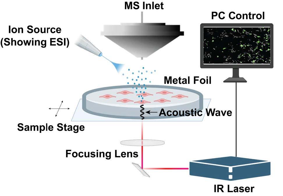
Matrix-assisted laser desorption/ionization (MALDI) has been the most commonly used method for MSI studied on a variety of samples, covering a reasonable range of molecules. However, limitations of MALDI MSI still exist, including low sensitivity toward small molecule metabolites with molecular weights lower than 400 Da, due to the interference and ion suppressions caused by extensive matrix ions, and low sensitivity toward less polar compounds. Besides, desorption and ionization occur simultaneously during a MALDI experiment, leading to in-source fragmentations of some labile compounds. Matrix application is also a major cause for the relatively low reproducibility of MALDI MSI experiments. Therefore, we are developing a gentler, matrix-free laser-based technique, laser-induced acoustic desorption (LIAD) to desorb intact molecules from the tissue surface, and couple it to other soft ionization techniques such as electrospray (ESI) and atmospheric pressure chemical ionization (APCI) for spatial omics studies.
LIAD employs a laser beam onto the back of a thin metal foil, generates acoustic waves transmitted through the metal foil, and knocks off molecules from the other surface of the foil gently into gas phase. Unlike laser desorption/ionization (LDI), little to no fragmentations are introduced to the desorbed molecules in LIAD experiments (Figure 1). We plan to apply LIAD as a new platform for MSI experiments to investigate spatial distributions of lipids and N-glycans of tissue sections. With this approach, we aim to obtain deeper understanding of metabolic pathways and alterations involved in various disease progression at molecular level.
We will conduct systematic studies by varying laser energy and number of laser shots, testing different metals and alloys and adjusting thicknesses of the metal foil to optimize the desorption efficiency and couple this LIAD MSI setup to different ion sources, so that both polar and nonpolar molecules can be imaged, providing a broad compound coverage.

2. High-confidence structural elucidation in MSI experiments
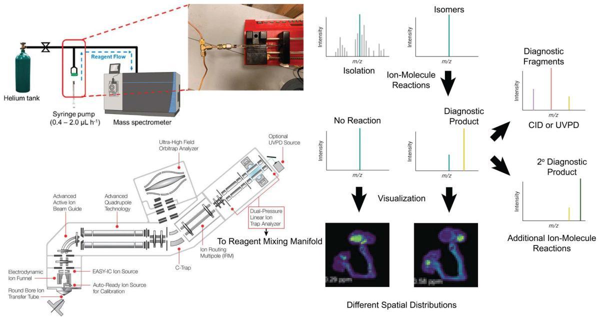
Structural elucidation in MSI experiments remains a major challenge. Functions of biomolecules such as lipids are highly dependent on their structures. Therefore, it is of great importance to obtain accurate structural information of biomolecules. Collision induced dissociation (CID) has been a widely used structural elucidation approach in many MS experiments. However, these tandem MS experiments cannot always provide adequate structural information. Other techniques, such as on-tissue derivatizations require sophisticated sample treatment, long reaction time and low reaction efficiency. Besides, current gas-phase reaction approaches such as ozonolysis or ion-ion reactions usually involve extensive instrumental development and modifications. We will conduct gas-phase ion-molecule reactions after molecules have been desorbed into gas phase from tissues or cells. These reactions provide diagnostic product ions or fragment ions upon further CID experiments, enabling structural determination of the molecules, especially isomers. Gas-phase ion-molecule reactions are usually highly efficient, whose reaction rate is mainly controlled by polarizability and dipole moments of the neutral reactant, and the charge state of the ionic reactant. Reactions usually complete within 50 ms, which matches the time scale of high-resolution MSI experiments. We will install a simplified reagent mixing manifold published previously to an Orbitrap mass spectrometer for neutral reagent introduction. The reagent mixing manifold will be connected to the dual-pressure linear ion trap of the mass spectrometer through the helium gas line, so that reaction products can either be transferred to the Orbitrap mass analyzer for high-resolution mass measurements, or subject to additional CID or secondary ion-molecule reactions in the linear ion trap. Spatial distributions of isomeric ions can be visualized distinctively, showing correlations of specific isomers to certain substructures of the tissue section (Figure 2).
We will couple this setup with either a desorption electrospray (DESI) MSI platform or the previously proposed LIAD MSI platform for experiments. The advantage of using the LIAD setup in these experiments is that LIAD separates desorption and ionization so that different ionization sources can be installed to cover a broader range of analytes. Furthermore, we will conduct mechanistic studies on identified diagnostic ion-molecule reactions to explore potential reaction mechanisms using DFT calculations, thus providing a theoretical basis for new reagent screening. We will test this setup using brain tissues provided by the Brain Institute, aiming to identify key lipids, N-glycans and other unidentified metabolites with high-confidence structural information that play pivotal roles in the development of Alzheimer’s disease.

3. Mechanistic studies of organic polyradicals toward small-molecule metabolites
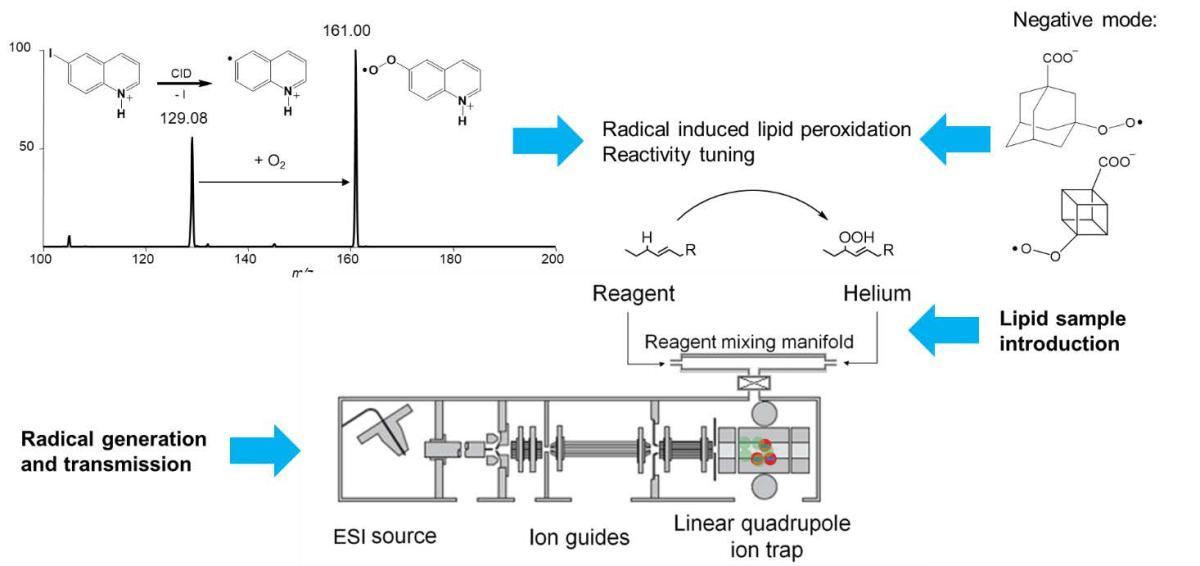
Organic polyradicals have shown high reactivity toward biomolecules such as oligonucleotides by abstraction of hydrogen atoms from nucleobases, leading to damage of the biomolecules. Some drugs have been designed based on this principle so that radicals are formed targeting at cancer cells. We will conduct ion-molecule reactions of organic polyradicals toward lipids, investigate reaction mechanisms, especially lipid peroxidations induced by oxyradicals generated in-situ in the gas phase (Figure 3), and explore and synthesize more new organic polyradicals that exhibit different reactivity toward biomolecules so that the cytotoxicity of the polyradicals can be monitored and tuned for better drug design process. The reactions of organic polyradicals and lipids will be studied using the aforementioned platforms for ion-molecule reactions. We will further investigate and regulate the radical-induced lipid peroxidations in cancer cells to mediate and control the cancer cell death and explore its potential as new cancer therapeutics.

References
- Zhu, H.; Kenttämaa, H. I. et al. J. Am. Soc. Mass Spectrom. 2016, 27, 1813–1823.
- Feng, E.; Fu, Y.; Ma, X.; Kotha, R.; Ding, D.; Kenttämaa, H. I., J. Am. Soc. Mass Spectrom. 2022, 33, 1794–1798.
- Widjaja, F.; Kenttämaa, H. I. et al. J. Am. Soc. Mass Spectrom. 2017, 28, 1392–1405.